
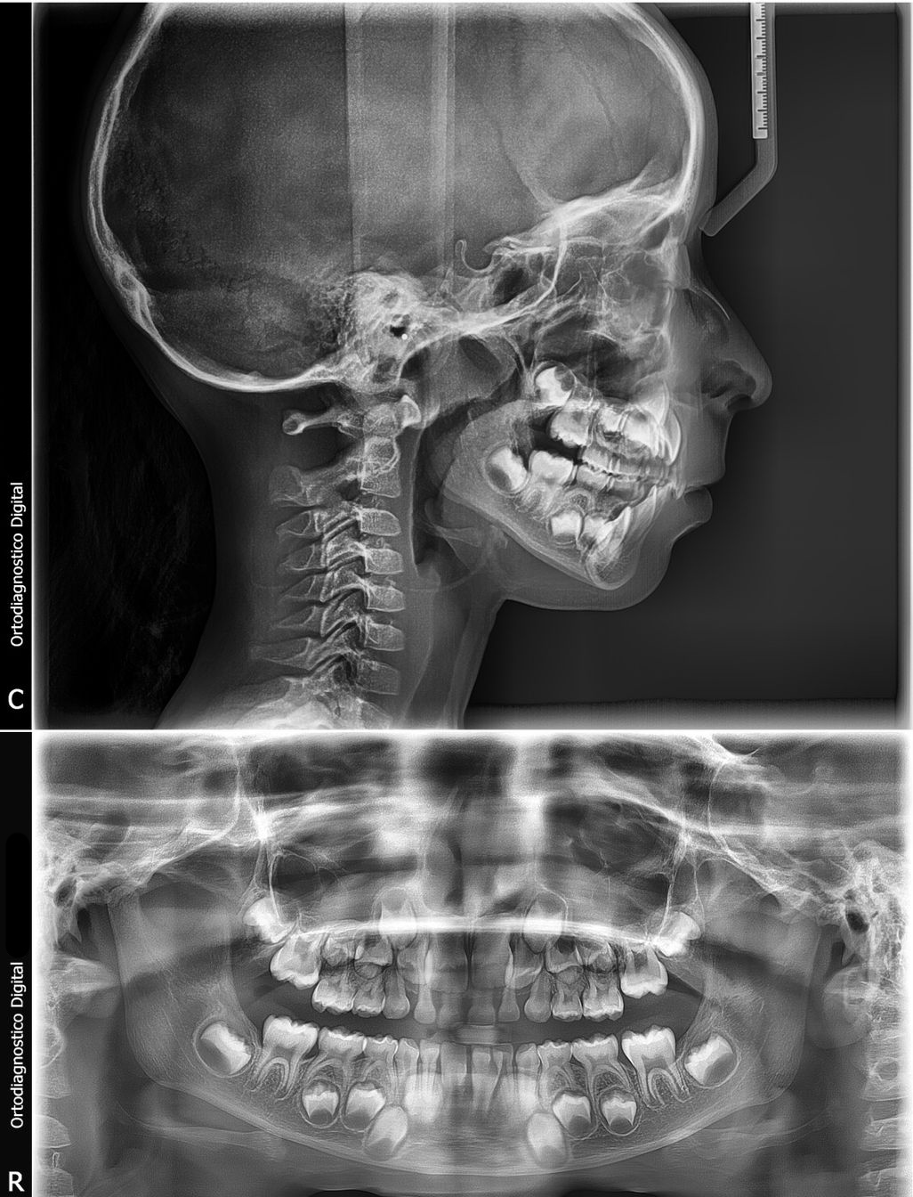
When your dentist asks you to get a dental X-ray, it's normal to have questions: what is it really for? Does it hurt? How much radiation does my body receive? Is it worth the cost? As a dentist with over two decades of practice in Monterrey, I understand. No one wants to undergo a study without understanding why it's necessary. In this article I'll explain — without jargon and with honesty — everything you need to know about dental X-rays so you can arrive at your appointment with peace of mind.
Dental X-rays are one of the most important diagnostic tools we have. It's not about "taking a picture for the sake of routine": every image answers a specific clinical question, and skipping it can lead us to miss hidden cavities, infections, or bone loss that are only visible thanks to dental X-rays. Let's go through, point by point, why they're indispensable, what types exist, and what you'll feel during the study.
What is a dental X-ray for? What your dentist sees that you can't
Dental X-rays are used to see what your eyes — and your dentist's — cannot observe during a clinical examination. Even with good lighting and mirrors in the dental chair, there are structures hidden between the teeth, below the gums, and inside the jawbone. That's where the radiographic image becomes irreplaceable.
These are the main reasons we request an X-ray:
- Interproximal cavities: cavities that form between two teeth, where floss passes but the dentist's mirror can't see. Only the X-ray reveals them at an early stage, when they're still reversible or easy to treat.
- Root and bone infections: periapical abscesses, cysts, and bone loss around the root are not visible to the naked eye. The X-ray shows bone density and the presence of dark lesions around the tooth.
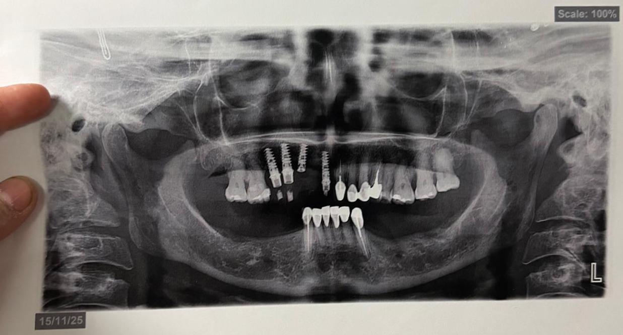
- Status of third molars: wisdom teeth that haven't erupted yet are often impacted or at dangerous angles. The panoramic X-ray tells us precisely their position relative to the inferior alveolar nerve.
- Treatment planning: dental implants, orthodontics, extractions, and root canal treatment require seeing the internal anatomy of the bone. A preliminary X-ray lets us design the procedure safely.
- Monitoring ultrasonic cleaning: when we perform prophylaxis with an ultrasonic scaler, the X-ray helps us assess how much subgingival calculus remains and confirm that the deep cleaning was effective.
In short, what dental X-rays are for: to accurately diagnose what can't be seen, to plan treatments with real data, and to track your progress over time. Without them, we'd be practicing dentistry blind.
Types of dental X-rays: panoramic, periapical, bitewing, and lateral
Not all X-rays are the same. Each type addresses a different clinical need, and in practice we combine several depending on the case. Understanding the types of dental X-rays helps you know what's being asked of you and why.
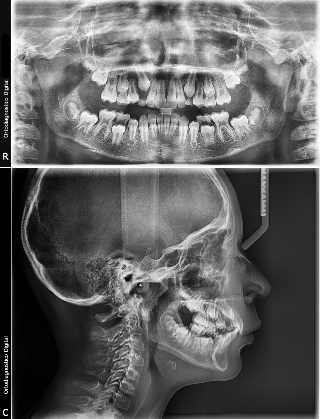
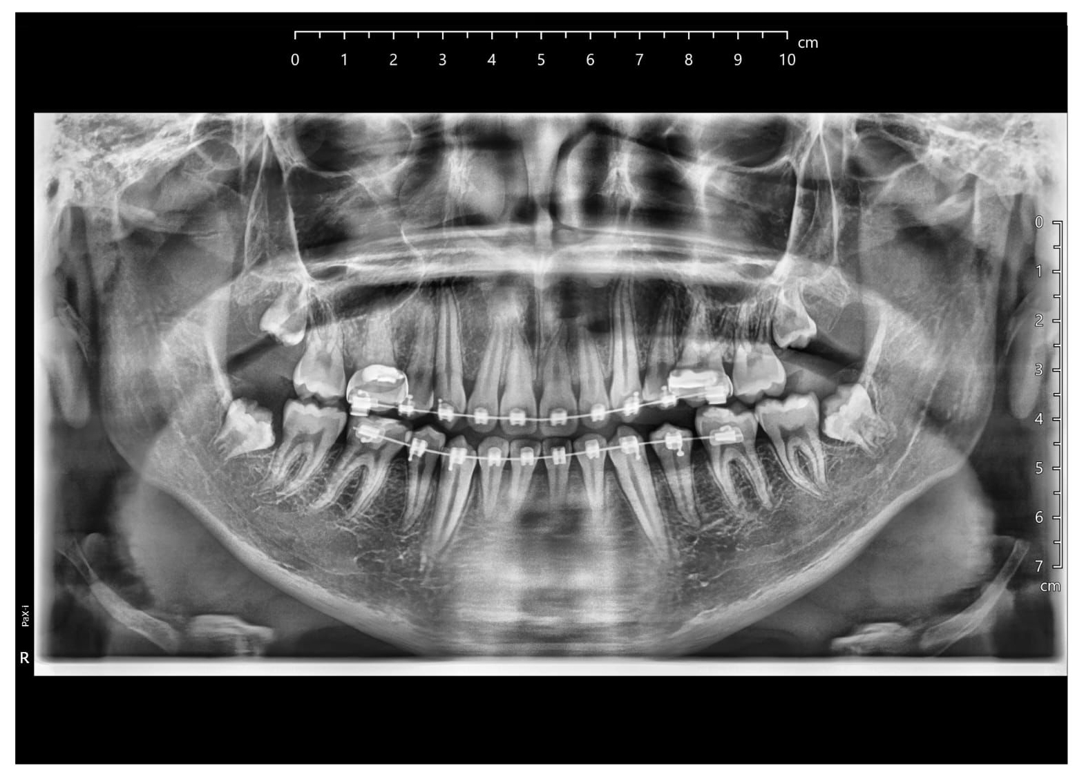
- Panoramic dental X-ray: a single image that shows the entire mouth at once: upper and lower teeth, upper and lower jaws, temporomandibular joint, and maxillary sinuses. It's taken from outside the mouth, standing in a machine that rotates around your head.
- Periapical X-ray: focuses on one or two complete teeth, from the crown to the tip of the root and the surrounding bone. This is the one we use for root canals, to evaluate abscesses, and to verify the seal of a filling.
- Bitewing X-ray: the patient bites down on a small tab and the sensor captures the crowns of the upper and lower teeth at the same time. It's ideal for detecting cavities between teeth and monitoring bone levels in patients with periodontal disease.
- Lateral X-ray (cephalometric): captures the profile of the head in a single projection. We order it in orthodontics and maxillofacial surgery to measure the position of the upper and lower jaws relative to the skull and to plan dental movements.
Each type uses a different dose of X-rays and is ordered at different points in treatment. Your dentist will choose the right combination based on your needs.
Panoramic dental X-ray: when it's ordered and what it reveals
The panoramic dental X-ray deserves its own section because it's the study we order most often on a first visit and during comprehensive check-ups. Why? Because in less than a minute you get a complete image of your mouth, and that gives us a very thorough clinical overview before touching a single tooth.
We order it in these cases:
- Initial assessment of a new patient: to see the overall condition of the entire mouth in a single image.
- Evaluating third molars (wisdom teeth): position, angulation, and proximity to the inferior alveolar nerve.
- Planning dental implants: evaluates the height and thickness of available bone for the implant screw, though for three-dimensional detail we order a CBCT scan.
- Orthodontics: to see how many permanent teeth still need to erupt and whether there are impacted teeth (upper canines, for example).
- Before maxillofacial surgery: fractures, cysts, odontogenic tumors, and bone lesions are evaluated first with a panoramic X-ray.
What does it reveal? Among other things: large cavities, bone loss from periodontitis, supernumerary teeth, missing teeth, root fragments, shape and position anomalies, and periapical lesions. It's not a fine-detail study for small cavities — for that we use the bitewing or periapical — but it is the best starting point.
In my practice at Centro Médico Obispado, I rarely begin a significant treatment without a recent panoramic X-ray. It's like getting a map before setting out on a trip: it saves you from surprises.
Does getting a dental X-ray hurt? What you'll actually feel
This is one of the most common questions in the office, and the short answer is: no, a dental X-ray does not hurt. But let me give you the long answer, because you deserve to know exactly what you'll feel.
Dental X-rays are non-invasive studies. There are no needles, no cuts, no instruments that touch your teeth in a painful way. All you have to do is hold the position the technician indicates:
- For a panoramic X-ray: you stand or sit in front of the machine, bite down on a small plastic tab to align your teeth, and the device rotates around your head for 15–20 seconds. You don't feel anything at all.
- For a periapical or bitewing: a small film or digital sensor is placed inside your mouth, against the tooth. You may feel the tab as a foreign object, and in some patients with a sensitive gag reflex it can cause mild discomfort when it touches the palate or tongue. The tab comes out in seconds.
- For a lateral (cephalometric): you stand against a head support and the X-ray is taken in a couple of seconds. There is no intraoral contact.
What can happen is that you may feel some discomfort from having to keep your mouth open or your head still, but that's not pain — it's posture. For patients with a sensitive gag reflex, there are techniques to minimize the response: smaller tabs, topical anesthesia on the palate, or simply taking more time. Always let us know; we will never force a study if you're feeling uncomfortable.
So if you were wondering "does a dental X-ray hurt?", you can relax: the study won't cause you pain. The most uncomfortable part is usually sitting and waiting for the file to process.
How much radiation does your body receive in a dental X-ray (and why you shouldn't worry)
This is the question I get most from parents in pediatric dentistry and from younger patients. And it's a serious question that deserves an honest, data-driven answer.
The radiation your body receives during a dental X-ray is very low. To put it in perspective, a panoramic X-ray is equivalent, in effective dose, to the background radiation you receive in approximately one day of normal life on Earth. In other words, if you sit in the sun and walk around Monterrey for a day, you've already received similar doses of natural radiation.
Some reference figures (effective dose, expressed in microsieverts, µSv):
- Digital bitewing: 2–5 µSv (the lowest dose).
- Digital periapical: 1–5 µSv.
- Digital panoramic: 10–20 µSv.
- Natural background radiation (one day in Mexico): ~8–10 µSv.
- Small-field cone beam CT (CBCT): 30–80 µSv.
- Medical chest CT scan: 1,500–2,000 µSv.
As you can see, a dental X-ray is well below any standard medical study, and most modern machines are now digital, which reduces the dose by up to 80% compared to traditional film. In addition, we use a lead apron with a thyroid collar for all intraoral studies.
Does this mean we take X-rays "just because"? No. The clinical principle is ALARA: As Low As Reasonably Achievable. We take the image only when the diagnostic benefit outweighs any risk, however small. If an X-ray won't change the treatment plan, we don't order it.
For pregnant women, the protocol is even more conservative: we defer non-urgent X-rays to the second or third trimester, and if they are essential, we use a double apron. The fetal dose from a panoramic X-ray is practically zero, but we prefer not to expose as a precautionary principle.
How much a dental X-ray costs and whether dental insurance covers it
Let's get to the point that matters most: your wallet. The cost of a dental X-ray in Monterrey varies depending on the type of study and the equipment used. Here's a general reference:
- Periapical or bitewing (single film): the most affordable option, within an accessible range for most patients.
- Digital panoramic: a mid-range cost; usually ordered once every 3–5 years or during initial assessments.
- Lateral cephalometric: mid-to-high cost, normally ordered together with the panoramic X-ray for orthodontics.
- Cone beam CT (CBCT): the most expensive, but we reserve it for cases where 3D imaging is essential (implants, complex surgery, endodontic retreatment).
It's important to note that at Especialidades Dentales (Centro Médico Obispado) we always give you a clear estimate before performing any study. There are no surprises in billing.
Do dental insurance plans cover them? In general, yes. Most dental insurance plans accepted in Monterrey include X-rays as part of the diagnostic workup. What varies is:
- Coverage frequency: some policies cover one panoramic X-ray per year, others every two or three years. Periapical and bitewing X-rays are typically covered more frequently.
- Reimbursement vs. direct payment: we help you review your policy and manage the necessary documentation.
- Annual limits: we check with you whether the study falls within the annual covered amount or whether it's better to defer it.
My recommendation: always bring your current insurance policy to your appointment and ask openly what's covered and what's not. Financial transparency is part of the respect we owe you.
X-rays in children: why pediatric dentistry needs them from an early age
Parents often ask me whether it's really necessary to take X-rays of children, especially if they're very young. And my answer is: yes, but with a protocol tailored to their age and needs. Modern pediatric dentistry works with clear criteria to minimize exposure without sacrificing diagnostic quality.
Why do we order X-rays in children?
- Detecting early cavities: children have thinner enamel on their primary teeth, and a cavity can progress quickly. The bitewing detects it before it starts to hurt.
- Monitoring eruption: the panoramic X-ray tells us whether permanent teeth are in the correct position, whether there are agenesis (teeth that never formed), or supernumerary (extra) teeth.
- Planning early orthodontics: the lateral and panoramic X-rays are the basis for deciding when to begin maxillary orthopedics treatment.
- Evaluating trauma: falls and bumps are common in childhood. A periapical X-ray confirms whether there's a root fracture or luxation.
How often? International clinical guidelines suggest a first bitewing X-ray when posterior teeth are in contact (around age 4–5) if the cavity risk is high, and a panoramic X-ray around age 6–7 to evaluate the transition from primary to permanent dentition. But this is a guideline, not a rigid rule: we evaluate each child individually.
Is the radiation safe for children? Yes, with digital technology and lead aprons with thyroid collars, the dose is minimal and the diagnostic benefits far outweigh the risk. Modern pediatric machines are calibrated to use the lowest possible dose.
My advice as a dentist (and as a person): don't be afraid to get an X-ray when your dentist recommends one, but don't be afraid to ask why they're ordering it either. A good professional will always explain the clinical reasoning. If they tell you "it's routine" and nothing more, ask further questions.
Practical summary
- Dental X-rays are used to see hidden cavities, infections, bone, third molars, and to plan treatments — they're not "a picture for the sake of routine."
- There are four main types: panoramic, periapical, bitewing, and lateral; each one answers a different clinical question.
- The panoramic X-ray is the first-choice study for comprehensive assessments and for planning implants, orthodontics, and surgery.
- It doesn't hurt: at worst, you feel an uncomfortable tab for a few seconds.
- Radiation is minimal, comparable to a day of natural sun exposure, and is further reduced with digital equipment.
- Cost varies by type; many dental insurance plans cover at least one panoramic X-ray per year.
- In children, X-rays are safe and necessary when there's a clinical reason that justifies them.
Did your dentist ask for an X-ray and you have questions?
At Especialidades Dentales (Centro Médico Obispado), Dr. Yadira Garza and Dr. Vanessa explain every image before taking it. Schedule your assessment via WhatsApp.
Book appointment via WhatsApp →References
- American Dental Association — Dental X-rays. Patient-facing information on dental radiographs and safety.
- FDA — Dental X-Ray Imaging. Regulatory guidance on radiation safety and equipment standards for dental imaging.
- ADA Science Institute — Dental Radiographs. Clinical guidelines on when to take dental X-rays and selection criteria.
- American Academy of Pediatric Dentistry — Radiograph Guidelines. Evidence-based recommendations on dental X-rays in pediatric patients.
- IAEA — Radiation Protection in Dentistry. International guidance on minimizing radiation exposure in dental practice.